Modular Raman Spectroscopy Kit

- 2.3 mm x 3.2 mm Coded Input Aperture
- 7.8 cm-1 to 9.7 cm-1 Spectral Resolution
- Typical Signal-to-Noise Ratio: 700:1
- Modular Design for Easy Customization
Application Idea
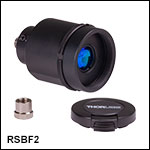
RSBF2 Fiber Input Adapter shown with an SMA-terminated multimode fiber patch cable and the RSB1(/M) Base Unit.
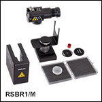
RSBR1(/M)
Reflective Front End Shown with the RSB1(/M) Base Unit (Items Sold Separately; Breadboard Not Shown)
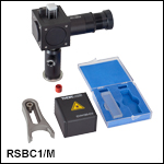
RSBC1(/M)
Cuvette Front End Shown with the RSB1(/M) Base Unit (Items Sold Separately; Breadboard Not Shown)

Please Wait
Building a Complete System from the Thorlabs Raman Spectroscopy Kit
A complete Raman spectroscopy system must include:
- 1 RSB1(/M) Spectrograph Base Unit
- 1 Front End for Collecting Raman Signal
- RSBR1(/M) Reflective Front End
- RSBC1(/M) Cuvette Front End
- Custom-Built Front End*
- Source with Excitation Between 680 nm and 785 nm (See Laser Requirements Tab; Sold Separately)
- PC for Running the Thorlabs OSA Raman Software (User Supplied)
*Custom front ends can be built using the RSBF2 Fiber Input Adapter with the RP35 Raman Fiber Probe or the RSBA1 60 mm Cage System Adapter.
Note: Do not mix imperial base units with metric front ends or vice versa. The input aperture height between imperial and metric units differs slightly due to the mounting posts.
| Key Specificationsa | |
|---|---|
| Input Aperture Size | 2.3 mm x 3.2 mm |
| Wavenumber Detection Range |
500 cm-1 to 1800 cm-1 @ 785 nm Excitation 2400 cm-1 to 3800 cm-1@ 680 nm Excitation |
| Spectral Resolution | <9.7 cm-1 at 500 cm-1 <7.8 cm-1 at 1800 cm-1 |
| Spectral Accuracy | <2.3 cm-1 at 500 cm-1 <1.8 cm-1 at 1800 cm-1 |
| Exposure Time: Typical Use Case | 1000 ms to 20 s |
| Signal-to-Noise Ratio (SNR) | 700:1 (Typical) >300:1 (Min) |
| Maximum Excitation Power | SM-Fiber: 250 mW MM-Fiber with Ø105 µm Core: 600 mW |
Features
- Modular, Large Field-of-View Raman Spectroscopy Kit with a Coded-Aperture Input
- Designed for 680 nm to 785 nm Excitation and an 815 nm to 915 nm Wavelength Detection Range
- Attached CMOS Monochrome Camera for Room Temperature Measurements
- Wavelength and Amplitude Calibrated for Comparison to Spectral Data Bases
- Optional Adapters for 60 mm Cage System Compatibility or Direct Fiber Input
- Spectra Recorded with Thorlabs' OSA Raman Software (See Software Tab)
Applications
- Quantitative Analysis of Chemical Mixtures
- Identification of Illegal or Dangerous Substances
- Quality Inspection (Pharmaceuticals, Food, etc.)
- Monitoring Chemical Processes at Production Sites
The Thorlabs Raman Spectroscopy Kit is a modular spectroscopy system that features a large (2.3 mm x 3.2 mm) coded aperture at the spectrograph input instead of a traditional slit to achieve a high signal-to-noise ratio (700:1), a low power density (at the sample), and room temperature operation. A pseudo-Hadamard mask of order 64, i.e., a defined pattern of elements for intensity modulation, is used at the input to increase the total light throughput of the system while maintaining high spectral resolution; please see the Coded Aperture tab for more information on coded-aperture (CODA) technology. This kit is designed for 680 nm to 785 nm excitation and an 815 nm to 915 nm detection range. A full list of specifications can be found on the Specs tab.
In comparison to traditional slit spectrographs, this kit averages Raman photons from a larger sampling volume, making it ideal for analyzing complex mixtures. The resulting output spectrum is a linear combination of the Raman fingerprints from the various molecules in the sampling volume, which can be identified through comparison to spectral databases.
A complete Raman spectroscopy system must include an RSB1(/M) base unit and a front end for collecting the scattered Raman signal; RSBR1(/M) and RSBC1(/M) front ends are sold below. A source with excitation between 680 nm to 785 nm, as well as a PC for running the Thorlabs OSA Raman software, must be supplied by the user. Thorlabs offers a Surface-Enhanced Raman Spectroscopy (SERS) Substrate (sold separately below) that can be used with the RSBR1(/M) front end to enhance the Raman signal by orders of magnitude. We also offer portable Raman spectrometers with integrated excitation lasers and automated substance analysis.
Base Units
The RSB1(/M) Base Unit, the core of Thorlabs' Raman spectroscopy system, is a volume-holographic grating imaging spectrograph that includes a coded input aperture and an attached Kiralux® monochrome CMOS camera. This unit is designed for 680 nm to 785 nm excitation. For excitation at 785 nm, we recommend using the FPV785M VHG-Stabilized Laser or any source that meets the specifications on the Laser Requirements tab. For easy comparison to spectral databases, the base unit is wavelength calibrated, as well as amplitude corrected using a NIST-certified polynomial during factory calibration.
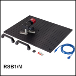
To get started instantly, this kit ships with the necessary accessories for mounting, including an aluminum base, mounting posts, and post clamps. A USB stick that contains a folder with the camera calibration dataset and a Microsoft Excel spreadsheet with macros for evaluating the excitation laser wavelength is also included. For A full list of items shipped with each base unit, please see the Shipping List tab. Note that a polystyrene calibration sample should be used to accurately determine the laser wavelength; these calibration samples are shipped with each front end or are available for purchase separately below.
Note: The attached Kiralux camera should not be removed; disassembly of the RSB1 base unit will destroy the system calibration.
Front Ends
This modular kit offers two front ends optimized for the collection of Raman-scattered photons from a variety of sample types. The RSBR1(/M) reflective front end is designed to examine powder and solid samples, while the RSBC1(/M) cuvette front end is ideal for liquids. Also available is the RSBF2 fiber input adapter, which couples Raman-scattered light carried via optical fiber from a user-designed setup to the base unit. For optical fiber excitation and light collection, we recommend our RP35 Bifurcated Reflection Probe for Raman Spectroscopy (sold below) which is compatible with the RSBF2 adapter. To build up a custom front end, the base unit is compatible with Thorlabs optomechanics through the SM1 (1.035"-40) and SM1.5 (1.535"-40) external threading on the aperture or our 60 mm cage system using the RSBA1 adapter (sold below).
Note that when paired with a user-supplied laser source, the RSBR1(/M) and RSBC1(/M) front ends and the RP35 Raman Fiber Probe provide a free space beam. Please consult your laser safety officer to ensure the safety of the setup before powering on any laser source. For further laser safety information, please refer to the user manual, which can be found by clicking on the red Docs icon (![]() ) next to each Item #.
) next to each Item #.
Note: RSBC1(/M) and RSBR1(/M) front ends built in 2021 or 2022 accommodate excitation only at 785 nm. Please contact Tech Support for assistance if you plan on adapting older front ends for excitation between 680 nm and 785 nm. Please be aware that while the wavelength detection range of the RSB1(/M) spectrograph will remain unchanged, the wavenumber detection range will change with the use of a different excitation wavelength.
Software
The Thorlabs OSA Raman Software is used to record Raman spectra, and the user-friendly software GUI allows the user to adjust the integration time of a single measurement, the analog gain, and the black level (pixel offset). The software also allows the user to easily change the axis units from wavelength to wavenumber, apply spectrum smoothing, and automatically correct the camera’s amplitude response with a NIST fluorescence standard for easy comparison to database spectra. For more information on the Raman software suite, please see the Software tab.
Raman Spectroscopy Kit Options
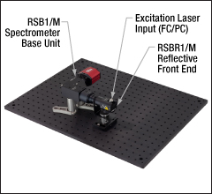
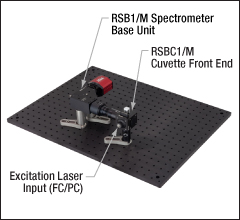
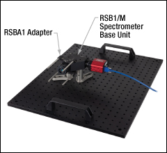
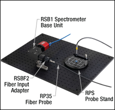
The images below show the possible setups that can be built from the Raman spectroscopy kit components. Each image depicts a spectrograph base unit mounted to an aluminum breadboard (included) with one of the available front ends or adapters. Note that the base unit, front ends, and adapters are sold separately. The Raman kit does not include an excitation source (see the Laser Requirements tab) or PC for running the available software.
Note: Do not mix imperial base units with metric front ends or vice versa. The input aperture height between imperial and metric units differs slightly due to the mounting posts.
Click to Enlarge
Raman Spectroscopy Kit Setup for Solid Samples
Click to Enlarge
Raman Spectroscopy Kit Setup for Samples in Cuvettes
Click to Enlarge
Raman Spectroscopy Kit Setup for Custom Builds
Click to Enlarge
Raman Spectroscopy Kit Setup for Fiber Input
Specifications
This tab describes the specifications for the base units and front ends, as well as specifications for the full kit.
Contents
- Raman Spectroscopy System Specifications
- Base Unit Specifications
- Reflective Front End Specifications
- Cuvette Front End Specifications
- Fiber Input Adapter Specifications
- Bifurcated Fiber Probe Specifications
| Raman Spectroscopy Kit Specificationsa | |
|---|---|
| Exposure Time: Technical Limitations | 0.036 ms (Min) to 22795 s (Maximum) in 0.022 ms Increments |
| Exposure Time: Typical Use Case | 1 s (Min) to 20 s (Maximum)b |
| Signal-to-Noise Ratio (SNR) | 700:1 (Typical) >300:1 (Minimum)c |
| Typical Amplitude Correction | NIST Standard (NIST SRM2241)d |
| Maximum Excitation Power | SM Fiber: 250 mW MM Fiber with Ø105 µm Core: 600 mWe, f, g |
Base Unit Specifications
All technical data are valid at 23 ± 5 °C and 45 ± 15% relative humidity (non-condensing).
| Item # | RSB1 | RSB1/M |
|---|---|---|
| Optical Specifications | ||
| Wavelength Detection Range | 815 nm to 915 nm | |
| Spectral Resolution (FWHM, Wavelength) | <0.65 nm | |
| Spectral Accuracy (Wavelength) | <0.15 nm | |
| Wavenumber Detection Range | 500 cm-1 to 1800 cm-1 (@ 785 nm Excitation) 2400 cm-1 to 3800 cm-1(@ 680 nm Excitation) |
|
| Spectral Resolution (FWHM, Wavenumber) | <9.7 cm-1 @ 500 cm-1 <7.8 cm-1 @ 1800 cm-1 |
|
| Spectral Accuracy (Wavenumber) | <2.3 cm-1 @ 500 cm-1 <1.8 cm-1 @ 1800 cm-1 |
|
| Beam Heighta | 3.25" (82.6 mm) |
84.1 mm (3.31") |
| Numerical Aperture | 0.22b | |
| Input Aperture | ||
| Type | Coded Aperture (Hadamard, 64th Oder) | |
| Aperture Material | Chromium on Fused Silica | |
| Aperture Size | 2.3 mm x 3.2 mm | |
| Single Element Size | 36 µm x 36 µm | |
| Grating | ||
| Type | Transmission | |
| Line Density | 1624 Lines/mm | |
| Center Wavelength | 871 nm | |
| Diffraction Efficiency (@ Center Wavelength, Average Polarization, and AOI = 45°) | >80% | |
| Camera/Sensor | ||
| Sensor Type | Monochrome CMOS, Non-Cooled | |
| Effective Number of Pixels (H x V) | 4096 x 2160 | |
| Imaging Area (H x V) | 14.131 mm x 7.452 mm (0.56" x 0.29") | |
| Pixel Size | 3.45 µm x 3.45 µm | |
| ADCc Resolution | 12 Bits | |
| General Specifications | ||
| Interface to PC | Hi-Speed USB 3.0 | |
| Dimensions (L x W x H) | 6.46" x 5.20" x 1.88" (164.2 mm x 132.0 mm x 47.6 mm) |
|
| Weight (Base Unit) | 0.867 kg | |
| Weight (Base Unit and Accessories) | 1.86 kg | |
| Ambient Operating Temperature | 10 °C to 40 °C (Non-Condensing) | |
| Storage Temperature | 0 °C to 55 °C | |
Reflective Front End Specifications
All technical data are valid at 23 ± 5 °C and 45 ± 15% relative humidity (non-condensing).
| Item #a | RSBR1 | RSBR1/M |
|---|---|---|
| Optical Specifications | ||
| Fiber Connector | FC/PC, Wide-Key | |
| Excitation Cleanup Filter, Bandpass | 680 nm to 785 nmb | |
| Longpass Filter, Cut-on Wavelength | 810 nmc | |
| Angle of Incidence | 30° | |
| Collection Geometry | Back-Scatteringd | |
| Maximum Excitation Power at Fiber Output | 250 mW SM Fiber 600 mW MM Fiber with 105 µm Coree,f,g |
|
| Focal Volumeh | ~10 mm3 | |
| General Specifications | ||
| Interface to the RSB1(/M) Base Unit | SM1 (1.035"-40) Internal Thread | |
| Dimensions (L x W x H)i | 3.00" x 3.89" x 4.36" (76.2 mm x 98.8 mm x 110.6 mm) |
75.0 mm x 99.1 mm x 114.6 mm (2.95" x 3.90" x 4.51") |
| Weightj | 0.5 kg | |
| Ambient Operating Temperature | 10 °C to 40 °C (Non-Condensing) | |
| Storage Temperature | 0 °C to 55 °C | |
Cuvette Front End Specifications
All technical data are valid at 23 ± 5 °C and 45 ± 15% relative humidity (non-condensing).
| Item #a | RSBC1 | RSBC1/M |
|---|---|---|
| Optical Specifications | ||
| Fiber Connector | FC/PC, Wide-Key | |
| Excitation Cleanup Filter, Bandpass | 680 nm to 785 nmb | |
| Longpass Filter, Cut-on Wavelength | 810 nmc | |
| Collection Geometry | 90°d | |
| Maximum Excitation Power at Fiber Output | 250 mW SM Fiber 600 mW MM Fiber with 105 µm Coree,f,g |
|
| General Specifications | ||
| Compatible Cuvette Type | 10 mm Light Path Dimensions: 12.5 mm x 12.5 mm x 45 mm (0.49" x 0.49" x 1.77") Optical Axis Height Above Cuvette Base: 8.5 mm (0.33") Four Polished Sides |
|
| Interface to the RSB1(/M) Base Unit | SM1 (1.035"-40) Internal Thread | |
| Dimensions (L x W x H)h | 2.66" x 4.54" x 5.19" (67.5 mm x 115.4 mm x 131.9 mm) |
69.3 mm x 115.2 mm x 133.4 mm (2.73" x 4.54" x 5.25") |
| Weight | 0.5 kg | |
| Ambient Operating Temperature | 10 °C to 40 °C (Non-Condensing) | |
| Storage Temperature | 0 °C to 55 °C | |
Fiber Input Adapter Specifications
All technical data are valid at 23 ± 5 °C and 45 ± 15% relative humidity (non-condensing).
| Item # | RSBF2 |
|---|---|
| Optical Specifications | |
| Wavelength Range | 650 nm to 1050 nm |
| Input Fiber Specifications | |
| Fiber Connector | SMA |
| Input Fiber Core Diameter (Max) | 2.5 mm |
| Numerical Aperture (Max) | 0.5 |
| General Specifications | |
| Device Compatibilitya | Exclusively Designed for the Thorlabs Modular Raman Spectroscopy Kit |
| Interface to RSB1(/M) Base Unit | SM1 (1.035"-40) Internal Thread |
| Dimensions | Ø31.8 mm x 48.9 mm (Ø1.25" x 1.93") |
| Weight | 0.055 kg |
| Ambient Operating Temperature | 10 °C to 40 °C (Non-Condensing) |
| Storage Temperature | 0 °C to 55 °C |
Bifurcated Fiber Probe Specifications
All technical data are valid at 23 ± 5 °C and 45 ± 15% relative humidity (non-condensing).
| Item # | RP35 | |
|---|---|---|
| Light Source Lega | ||
| Fiber Connector | FC/PC Narrow Key | |
| Wavelength Range | 680 ± 0.5 nm to 785 ± 0.5 nm | |
| Maximum Input Power | 300 mW @ 680 nm 600 mW @ 785 nm |
|
| Fiber Core Diameter | Ø200 ± 4 μm | |
| Fiber Numerical Aperture | 0.22 ± 0.02 | |
| Spectrometer Lega | ||
| Fiber Connector | SMA905 | |
| Wavelength Range | 815 nm to 950 nmb | |
| Number of Fibers | ≥65 Fibers | |
| Illuminated Area | 2.1 mm (0.08") | |
| Individual Fiber Core Diameter | Ø200 ± 4 μm | |
| Individual Fiber Numerical Aperture | 0.22 ± 0.02 | |
| Material Specifications | ||
| Fiber Cores | Low-OH Fused Silica | |
| Furcation Tubing | Sheathed Stainless Steel | |
| Lensc | Fused Silica | |
| Operating Conditions | ||
| Operating Temperature Range | 5 °C to 35 °C (Non-Condensing) | |
| Storage Temperature Range | -40 °C to 70 °C | |
| Mechanical Specifications | ||
| Probe Tip Diameter | Ø6.35 mm (1/4") | |
| Largest Dimension of Combined Legs | 9.5 mm x 18.4 mm (0.38" x 0.73") | |
| Minimum Bending Radius | 80 mm (3.15") | |
| Length | Individual Leg | 1 ± 0.075 m (39.4 ± 3.0") |
| Overall | 2 +0.075 /-0 m (78.8 +3.0/-0") | |
| Weight | 0.2 kg (0.44 lbs) | |
| Laser Requirements | |
|---|---|
| Wavelength | 680.0 ± 5.0 nm to 785.0 ± 0.6 nm |
| Spectral Bandwidth (FWHM) | <0.15 nm |
| Wavelength Stabilization | <0.02 nm |
| Thermal Stabilization | ±0.1 °C (Max) (Wavelength Stability: <0.02 nm) |
| Output Power (Max) | 600 mWa |
| Recommended Output Power (Approximate) | 250 mW ± 150 mW |
| Fiber Connector | FC/PC |
| Fiber Core Diameter | Ø105 µm |
| Fiber NA | 0.22 |
Recommended Laser for the Raman Spectroscopy System
Raman spectroscopy requires a laser source with a narrowband, stabilized output to produce spectra with clean signatures. Because the number of Raman-scattered photons is linearly proportional to the intensity of the excitation source, the laser should also have a high output power. The laser specifications required for proper operation of Thorlabs' Raman system are shown in the table to the right. Note that, as the sample needs to be illuminated homogeneously, a Ø100 μm to Ø200 μm multimode fiber should be used to guarantee a flat-top profile on the sample after collimation by the front end.
For 785 nm excitation, we recommend using Thorlabs' FPV785M Volume-Holographic-Grating- (VHG) Stabilized Laser. This laser uses a volume holographic grating to provide narrow-linewidth operation at 785 nm, and it is pigtailed to an FC/PC-terminated multimode fiber to accommodate high-power, homogenous sample illumination. To achieve a wavelength-stable output, we also recommend using this laser with our CLD1015 Laser Driver and Temperature Controller.
The use of armored multimode fiber patch cables with a >Ø105 µm core is strongly recommended even if the laser is fiber pigtailed. Directly connecting to the front end with a non-armored fiber increases the risk of exposure to dangerous radiation in case the fiber becomes damaged.
Note that when paired with a user-supplied laser source, the front ends provide a free space beam. Necessary laser safety protocols will depend on the user's chosen laser source and the lab environment. Please consult your laser safety officer to ensure the safety of the setup before powering on any laser source.
| Input Fiber Requirements | |
|---|---|
| Fiber Connector to RSBR1(/M) or RSBC1(/M) Front Ends | FC/PC |
| Fiber Type | Multimode Fiber |
| Fiber Core Diameter | Ø100 µm (Minimum) Ø105 µm (Typical) Ø200 µm (Maximum) |
| Fiber NA | 0.22 |
| Recommended Fiber | MR16L01 Armored Fiber Patch Cable |
Fiber Specifications for the Raman Spectroscopy System Front Ends
The table to the right shows the specifications required for the fiber that connects the excitation laser to the reflective and cuvette front ends. For the RSBF2 Fiber Adapter, we recommend using our RP35 Reflection Probe for Raman Spectroscopy.
| OSA Raman Software Recommended System Requirements | |
|---|---|
| Operating System | Windows® 10 (32-Bit, 64-Bit) |
| Processor (CPU) | Intel Pentium 4 or AMD Athlon 64 3000+ |
| Memory (RAM) | 2.0 GB |
| Graphic Card Resolution (Min) |
800 x 600 Pixels |
| Hard Drive (Min) | 2 GB of Available Disk Space (32-Bit, 64-Bit) |
| Interface | Free High-Speed USB 3.0 Port |
| ThorCam Recommended System Requirements | |
|---|---|
| Operating System | Windows® 7, 10, or 11 (64 Bit) |
| Processor (CPU)a | ≥3.0 GHz Intel Core (i5 or Higher) |
| Memory (RAM) | ≥8 GB |
| Hard Driveb | ≥500 GB (SATA) Solid State Drive (SSD) |
| Graphics Cardc | Dedicated Adapter with ≥256 MB RAM |
| Motherboard | USB 3.0 (-USB) Cameras: Integrated Intel USB 3.0 Controller or One Unused PCIe x1 Slot (for Item # USB3-PCIE) GigE (-GE) Cameras: One Unused PCIe x1 Slot |
| Connectivity | USB or Internet Connectivity for Driver Installation |
Software for Raman Spectroscopy Kit
Thorlabs' Raman Spectroscopy Kit is controlled by our Thorlabs OSA Raman software package. This software features a straightforward, intuitive, responsive interface that exposes all functions in one or two clicks. Click the software link below (left) to download the latest version of the OSA Raman Software package.
The alignment of the RSBF2 Fiber Input Adapter can be monitored with the ThorCam™ Software, which is an image acquisition software package designed for use with our scientific cameras on 32- and 64-bit Windows® 7 or 10 systems. The intuitive, easy-to-use graphical interface provides camera control as well as the ability to acquire and play back images. Click the software link below (right) to download the latest version of the ThorCam Software package.
Software
OSA Raman Version 3.0
Includes a GUI for acquiring spectra with the RSB1(/M) Base Unit from the Raman Spectroscopy kit.
Software
ThorCam™ Version 3.7.0
The ThorCam software is used in the alignment procedure for the RSBF2 Fiber Input Adapter. Click the button below to visit the ThorCam software page.
Click to Enlarge
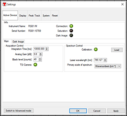
The integration time, analog gain, and black level can be adjusted in the settings dialog. Also in this window are the device status icons and the calibration information.
Click to Enlarge
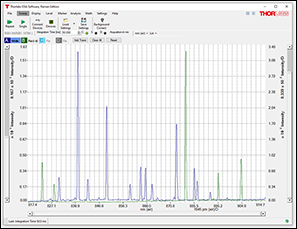
The OSA Raman software can display data on the primary and secondary vertical axes, which are on the left and right of the graph, respectively. A trace displayed on the secondary axis will have a small arrow displayed next to its icon in the Trace controls bar. The software can handle up to 26 traces.
OSA Raman Software Highlights
The text below summarizes several key features of the OSA Raman software. Complete details on the software are available in the manual (PDF link).
Settings Dialog
The OSA Raman software allows critical parameters for Raman spectroscopy to be adjusted, including the integration time, analog gain, and black level. Also included in the device settings are indicator buttons to show whether the camera is connected to the PC, the saturation state of the camera, and whether acquisition of a new dark image is required. Spectra units can also be set to either wavelength (nm) or wavenumbers (cm-1).
Built-In Tools for Analysis
Robust graph manipulation tools include automatic and manual scaling of the displayed portion of the trace and markers for determining exact data values and visualizing data boundaries. Automated peak and valley tracking tools identify up to 2048 peaks or valleys within a user-defined wavelength range and follow them during continuous spectrum acquisition. Statistical parameters of traces such as standard deviations, RMS values, and weighted averages are available, and a curve fit tool fits polynomial, Gaussian, and Lorentzian functions to the spectrum.
To compare acquired spectra to Raman signatures of known substances, spectra from external databases can be be imported into the software using CSV files.
Data Export
The Raman trace can be saved in the OSA Raman software spectrum file format (.spf2x) or exported into various file formats, including Matlab, Galactic SPC, JCAMP-DX, CSV, and text formats. Note that the OSA Raman software can load but not export .spf2 file formats generated with other Thorlabs OSA software.
Shipping List
The following are shipped with the RSB1(/M) Base Unit:
- Base Unit for Raman Spectroscopy with Protective SM1 Cap
- USB 3.0 USB Cable (Micro B to A) to Connect the Camera to the PC
- MB1824 (MB4560/M) 18" x 24" x 1/2" (450 mm x 600 mm x 12.7 mm) Aluminum Breadboard
- BBH1 Breadboard Lifting Handles
- 2 x RS2.5P(/M) Ø1" (Ø25.0 mm) Mounting Posts, L = 2.5" (65 mm)
- 2 x CF125C(/M) Clamping Forks
- USB Stick with Coded Matrix Calibration Data and Microsoft Excel Spreadsheet with Macros for Evaluating the Excitation Laser Wavelength
- Quick Reference
The following are shipped with the RSBR1(/M) Front End for Reflective Measurements:
- Front End for Reflective Measurements with Protective SM1 and FC/PC Caps
- Sample Table with a BA2F(/M) Mounting Base
- RPC Polystyrene Calibration Chip
- Stray Light Shield
- Quick Reference
The following are shipped with the RSBC1(/M) Front End for Cuvette Measurements:
- Front End for Cuvette Measurements with Protective SM1 and FC/PC Caps
- TR1.5 (TR40/M) Ø1/2" (Ø12.7 mm) Mounting Post, L = 1.5" (40 mm)
- PH2E (PH50E/M) Ø1/2" (Ø12.7 mm) Pedestal Post Holder
- CF125C(/M) Clamping Fork
- 3500 µL Macro Fluorescence Synthetic Quartz Glass Cuvette1
- RBP Polystyrene Block for Calibration
- Quick Reference
The following are shipped with the RSBF2 Fiber Input Adapter:
- Fiber Input Adapter with Protective SM1 and SMA Caps
- Quick Reference
The following are shipped with the RP35 Raman Fiber Probe:
- Bifurcated Fiber Probe with Plastic Protective Caps for SMA, FC/PC, and Fiber Probe Ends
- Quick Reference
The following are shipped with the RSBA1 Adapter for 60 mm Cage Systems:
- 2 Mounting Brackets
- 4 8-32 (9/64" Hex) and 4 M4 (3 mm Hex) Cap Screws
- Quick Reference
- The CV10Q35F Macro Fluorescence Cuvette is an equivalent replacement.
Click to Enlarge
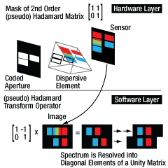
A spectrograph with a coded aperture uses a slit pattern to increase light throughput while maintaining high spectral resolution. This example shows a Hadamard mask of order 2, while the mask used in the RSB1(/M) base unit is order 64.
Click to Enlarge
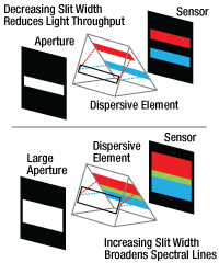
Traditional imaging spectrographs require narrow slits to achieve high spectral resolution. This limits the light that can pass through the spectrograph.
Coded-Aperture (CODA) Raman Spectroscopy
The Thorlabs Raman Spectroscopy System is based on an imaging spectrograph that features a diffraction grating as the dispersive element. Traditional spectrographs of this design face a trade-off between the desired spectral resolution and the achievable light throughput. This is because the dispersive element produces spectrally separable images of the same slit image. When the slit is wider, the light throughput may be increased, but the spectra may no longer be separable, leading to loss of the spectral information.
In contrast, the Thorlabs' Raman system spectrograph replaces the single-slit input aperture of conventional systems with a defined pattern of multiple slits, which is known as a coded-aperture (CODA) input. This spatial amplitude modulation mask acts as a convolution operator with an orthogonal basis in two dimensions. The resulting mesh image from the sensor is reconstructed using the inverse transform operator. This encoding uses the complete large window of the mask for optical input, while the output spectral resolution is defined by dimensions of a single mask element.
The pure math engine of a Hadamard transform is adopted for the Raman spectroscopy application; please see the reference article for more details1. Hadamard matrices consist of elements that only take values of 1 and -1. Since the -1 element cannot be realized in a real-world mask, our pseudo-Hadamard optical mask maps the -1 to 1 values to intensity modulations from the range of 0 (light blocked) and 1 (pixel transparent). The necessary spectrum reconstruction algorithms are implemented in the supplied Thorlabs OSA Software, as well as corrections for the unavoidable optical distortions due to a large input field. Device specific calibration data for compensation of the exact instrument distortions and transmission characteristics are shipped on a USB with each RSB1(/M) base unit.
References
- M.E. Ghem et al., "Static Two-Dimensional Aperture Coding for Multimodal, Multiplex Spectroscopy," Applied Optics, vol. 45, no. 13, 2006.
Polarization-Dependent Raman Spectroscopy
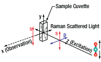
Click to Enlarge
Coordinate Axes for Polarization Orientation
Controlling the polarization of the excitation and Raman-scattered light can provide information about the symmetry of bond vibrations within a molecule. In the as-delivered configuration (90° geometry), the RSBC1(/M) cuvette front end only detects s-polarized light unless the excitation light is completely unpolarized. The signal strength of peaks resulting from symmetrical vibrational modes can be optimized by rotating the collimator or by adding polarization optics to the excitation and collection paths. Note that due to its 180° geometry, the RSBR1(/M) reflective front end is not sensitive to polarization.
When adjusting any part of the Raman spectroscopy kit, carefully follow laser safety instructions for the user-supplied laser; a collimated laser beam is emitted from the fiber port of the RSBC1 front end and collimator bundle when the user-supplied laser is connected. Any laser should be turned off before modifying the setup.
Background
The symmetry of the bond vibration behind a Raman peak is characterized by the depolarization ratio:

where I|| and I⊥ are the peak intensities for parallel and perpendicular polarization of the observed Raman scattered light with respect to the excitation light. A vibrational mode is completely symmetric and referred to as a polarized band when ρ < 0.75. When ρ ≥ 0.75, a vibrational mode is not completely symmetric and is called a depolarized band.
In this application note, excitation light propagates along the z-axis, s-polarized means light polarized in the y-direction, and p-polarized means polarization in the x-direction.
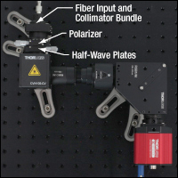
Click to Enlarge
Excitation and Collection Paths with Polarization Optics Included
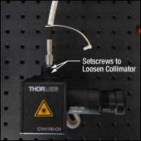
Click to Enlarge
Loosen Two 1/6" (1.5 mm) Setscrews to Rotate the Collimator
Optimizing Raman Signal via Collimator Rotation
In general, excitation light is not completely non-polarized, as the original polarization of the laser source is partially preserved even after passage through a considerable length of multimode fiber. The setup consisting of the RSB1(/M) base unit and RSBC1(/M) cuvette front end only allows for the observation of s-polarized light. The ratio of parallel and perpendicular components contributing to the measured Raman spectrum changes upon rotation of the fiber around the z-axis, which can strongly enhance or attenuate the Raman peaks.
It is not possible to rotate the fiber within the collimator FC/PC fiber port, and fiber rotation around the z-axis can only be achieved by rotating the collimator with the fiber attached to it. To rotate the collimator, loosen the two 1/16" (1.5 mm) nylon-tipped screws that secure it in the fiber input.
When adjusting any part of the Raman spectroscopy kit, carefully follow laser safety instructions for the user-supplied laser; a collimated laser beam is emitted from the fiber port of the RSBC1 front end and collimator bundle when the user-supplied laser is connected. Any laser should be turned off before modifying the setup.
Including Polarization Optics in the Excitation and Collection Path
To accurately determine depolarization ratios, polarization optics can be added to the excitation and collection paths of the Thorlabs Raman system that includes the RSB1(/M) base unit and RSBC1(/M) cuvette front end.
The excitation path can be modified by unscrewing the fiber input and collimator bundle from the front end and mounting them in an optics mount, such as CP33(/M). Then, a half-wave plate and polarizer, mounted in appropriate optics mounts, can be placed in front of the input port of the main body of the front end. The collection path can be modified by unscrewing the baffle assembly from the base unit and inserting a half-waveplate and polarizer. Note that this construction must be made light tight and samples that have increased scattering should have user-supplied baffles to help prevent unwanted straylight from entering the spectrograph.
Note: P-polarized Raman-scattered light is observable in a 180° geometry, i.e. transmission setup. While it is possible to convert the RSBC1(/M) cuvette front end into a 180° setup and observe Raman signal in this configuration, a significant amount of excitation light will reach the camera sensor. This leads to increased background and noise levels in the spectral range of the base unit. In this modified orientation, it is recommended to use additional filters, such as the FELH0800, to supress the excitation light in the Raman-scattered light path.
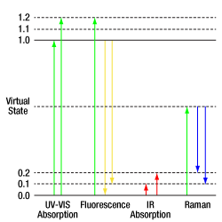
Click to Enlarge
Energy Levels of Various Radiation Types
Raman Spectroscopy
Raman Spectroscopy is a well-established technique for the characterization of chemical substances in solid, powder, liquid, or gas forms. It is based on the detection and analysis of Raman scattered light emitted from a sample upon exposure to monochromatic light.
Raman Scattering
Atoms in a molecule are held together by chemical bonds. Depending on the number of atoms and the geometry of the bonds, there are different ways molecules can vibrate. Each unique mode of vibration can be excited only with a certain, well defined amount of energy. When an exciting photon is absorbed by the molecule, it may excite a vibrational mode or absorb energy from an existing vibrational mode before being re-emitted. The loss or gain of energy due to this interaction with a vibrational mode means the photon is inelastically scattered, and this interaction between the light and the material is called the Raman effect. The resulting shift in energy and wavelength of the scattered photon due to this inelastic scattering (Raman scattering) is called the Raman shift. Raman spectroscopy measures this Raman shift.
Raman scattering is a rather rare event and occurs for only about 1 in 1 million exciting photons. It is, therefore, critical to excite the sample with a high-power light source and to collect as much Raman-scattered light as possible for analysis. To generate sufficient Raman scattering, the sample is typically exposed to high-intensity monochromatic laser light.
Because excitation of a whole set of vibrational states of a sample with a defined amount of energy (the exciting laser light) results in material-specific Raman scattering, a measured Raman spectrum may be considered a chemical fingerprint of the substance. The vibrations detected by Raman spectroscopy may differ from those modulating the molecular dipoles, which are observable by IR spectroscopy, and make it a valuable source for material analysis and complementary to other types of vibrational spectroscopy.
Energy Shift Due to Inelastic Scattering
Raman spectroscopy analyzes the shift in energy due to inelastic scattering caused by the Raman effect. The wavelength of the inelastically scattered photon is related to the excitation wavelength through the following energy equation describing the Raman scattering event:

where Evib is the vibrational energy of the molecule affected by the exciting photon, Eout is the energy of the scattered photon, and Ein is the energy of the exciting photon.
The photon's energy and frequency are related via the Planck-Einstein relation:

with h = 6.6260715 x 10⁻³⁴ J⋅s, fin is the incoming photon frequency in Hertz. The analogous relation holds for the scattered photon (Eout = hfout).
Using equations 1 and 2, as well as λ = c/f where c is the speed of light, the Raman shift of a molecular vibration can be characterized with knowledge of the precise wavelength of the excitation wavelength λin based on the following equation:

Describing Raman scattering in wavelength space is a disadvantage, as it depends on the precise excitation wavelength. However, it is fixed in the Raman-shift scale, or wavenumber scale, in units of cm-1. Therefore, in order to compare Raman spectra of a substance between different experimental setups, Raman spectra are represented as 2D plots of measured relative intensity (Y-axis) in Raman shift of the inelastic scattered light in wavenumbers (cm-1, X-axis). This unit is, in contrast to nanometers, a scale proportional to energy.
| Posted Comments: | |
Jiwon Yune
(posted 2023-08-23 13:43:30.833) I am having a difficult time installing the Thorlabs Raman software. Although .NET framework v4.7.2 and above are installed on my PC, the installer tells me that .NET framework is missing, when I run it for the first time. FYI, I am using Windows 11 on Intel. How can I fix this issue? soswald
(posted 2023-08-24 03:46:15.0) Dear Jiwon Yune,
thank you for your feedback. Can you please try installing .NET framework version 4.7.2 or 4.8 from here: https://dotnet.microsoft.com/en-us/download/dotnet-framework/net472
I have reached out to you directly to further assist with troubleshooting. NICOLAS DAUGEY
(posted 2023-06-08 16:35:06.01) Dear all,
I just found this Raman spectrometer. I am very interested by your spectrometer especially with your high throughput special slit and the choice of two lasers.
Is it recent product from your side?
Is it possible to test it in a comparative measurement at our lab with another spectrometer?
Thanks in advance,
N. Daugey soswald
(posted 2023-06-09 02:16:06.0) Dear Nicolas,
thank you for your feedback. Both our modular Raman spectroscopy kit as well as the portable Raman spectrometers (RASP1/RASP2) were indeed added to the catalogue quite recently.
We are happy to provide loan devices so you can test them at your lab. I have reached out to you directly to discuss this further. Carles Ros
(posted 2022-07-04 09:34:40.42) Hi, we want to set a similar setup to the Raman spectroscopy kit and have few questions:
- you propose the FPV785M laser, and to adapt it to the system with a Armored Multimode Fiber Optic Patch Cable. how we connect the laser, which already comes with a fibre, with the armored fibre? or can it be substituted?
- which of the armored fibers is better and which is the exact difference? M29L o M93L are the ones you suggest.
- this raman setup is only possible with the 785nm laser? could it be set with a shorter wavelength light?
thanks a lot! nreusch
(posted 2022-07-07 03:45:18.0) Thank you for contacting us. If you use an excitation laser that is pigtailed with a fiber which is not armored, we recommend not connecting this comparatively fragile fiber to the front end directly due to the risk of exposure to dangerous radiation in case the fiber gets damaged. You could use a mating sleeve to connect the non-armored fiber to an armored fiber.
Comparing e.g. M93L01 and M29L01, M93L01 comes with a 1500 µm core diameter with FT05SS tubing, whereas e.g. M29L01 is a 600 µm core diameter patch cable with FT030 tubing. This means the fiber type as well as the tubing differs. Both have SMA connectors that would not match the input port of the kit. The input fiber requirements are listed under tab “Laser Requirements”. You would need a multimode fiber patch cable with FC/PC connector and a core diameter between 100 µm (minimum) and 200 µm (maximum) and a numerical aperture of 0.22. The recommended fiber would be MR16L01, i.e. an armored 105 µm core diameter fiber with FC/PC connectors. As alternative, you could use one of the 200 µm FC/PC patch cables that are listed at https://www.thorlabs.com/newgrouppage9.cfm?objectgroup_id=5684.
If you want to use the Raman bundle in the high-wavenumber region between 2800 cm^-1 and 3800 cm^-1, you could choose an excitation laser at 680 nm, but you would also need to change the filter in the front end of the kit. I will contact you to discuss further details about your application. |

Must be Purchased Separately
- One of the Following Front Ends:
- Source with Excitation between 680 nm and 785 nm (See Laser Requirements Tab)
- PC for Running the Thorlabs OSA Raman Software
For the full list of components shipped with this base unit, please see the Shipping List tab.
- Volume-Holographic Grating Imaging Spectrograph with a 2.3 mm x 3.2 mm Coded Aperture
- Attached and Aligned Monochrome CMOS Camera with a USB 3.0 Interface (Item # CS895MU)*
- Designed for 680 nm to 785 nm Excitation Wavelength
- Amplitude Corrected using a NIST-Certified Polynomial (NIST SRM2241)
- Includes USB Dive with Camera Calibration Data
The RSB1(/M) Base Unit is the core of Thorlabs' Raman Spectroscopy Kit, which is a modular system ideal for detecting low-intensity Raman signal. It is designed for a laser with excitation between 680 nm and 785 nm. Spectra are recorded in the 815 nm to 915 nm wavelength range which, when using a 785 nm excitation laser, corresponds to 500 cm-1 to 1800 cm-1. Each base unit includes an imaging spectrograph with a volume-holographic grating, a large coded-aperture (CODA) input, and an attached Kiralux Monochrome CMOS Camera; the grating and camera have wavelength-dependent efficiencies that are amplitude-corrected using a certified NIST standard during factory calibration. For optimal extraction of the Raman-scattered light, this base unit uses an internal longpass 810 nm filter.
Click to Enlarge
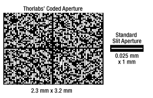
Size comparison of the coded aperture and a standard slit aperture. The coded aperture is a pseudo-Hadamard mask of order 64; the white and dark areas represent where light is transmitted through the mask or blocked, respectively.
To assemble a full Raman spectroscopy system, this base unit must be equipped with a front end (see below), a source with excitation between 680 nm and 785 nm, and a PC. For excitation at 785 nm, we recommend using Thorlabs' FPV785M VHG-Stabilized 785 nm Laser as the excitation source; however, a full list of required specifications for alternative excitation sources can be found on the Laser Requirements tab.
Each base unit includes an MB1824 (MB4560/M) 18" x 24" x 1/2" (450 mm x 600 mm x 12.7 mm) aluminum breadboard; see the Shipping List tab for a full list of items shipped with the base unit. To mount to the breadboard, the spectrograph housing features three 1/4"-20 (M6 x 1) taps for RS2.5P(/M) mounting posts. Note that this device is isometric and can be mounted upside down using three additional mounting taps located on the top of the housing.
*Note that the attached Kiralux camera should not be removed; disassembly of the RSB1 base unit will destroy the system calibration.
Coded Input Aperture
This base unit utilizes a pseudo-Hadamard mask of order 64 as the aperture, instead of a single slit like conventional spectrographs. With an aperture size of 2.3 mm x 3.2 mm, this spatial amplitude modulation mask improves light-throughput, or the system étendue, allowing enough Raman-scattered light to reach the CMOS sensor for room temperature operation.
Increased étendue of the spectrograph's input also enables a larger spot size (Ø1 to Ø2 mm), which allows sample excitation with an unfocused laser beam. This is ideal for nonhomogeneous samples, such as powders and pills, that require an average over a large area to determine composition. An unfocused excitation beam also results in a lower power density, reducing the risk of laser-induced damage to the sample. For more information on CODA technology, please see the Coded Aperture tab.

Click for Details
The OSA Raman Software GUI allows for easy acquisition of Raman spectra, and it includes built-in tools for analysis. Shown here is a polystyrene spectrum where peak information has been identified with the peak track tool.
Mounting to Front Ends and Adapters
The Raman base unit features an SM1 (1.035"-40) external thread adapter for mounting the RSBR1 and RSBC1 front ends or the RSBF2 fiber input adapter onto the input aperture. To facilitate excitation and scattered light collection, the RP35 Raman Fiber Probe connects directly to the RSBF2 adapter. For customer-designed front ends, the base unit also includes an SM1.5 (1.535"-40) external thread adapter, which can be used, for example, with our Ø1.5" lens tubes. Alternatively, the base unit has two 8-32 (M4 x 0.7) taps for the RSBA1 adapter sold below; this adapter can be used to provide compatibility with Thorlabs' 60 mm cage system components.
Amplitude Correction
The components of this spectrograph, such the grating and detector, have wavelength-dependent efficiencies. To compensate for this, these spectrographs are amplitude corrected using a NIST SRM2241 certified standard during factory calibration, which ensures compatibility with spectral databases. This standard defines a polynomial with relative, quantitative fluorescence intensity for the base unit detection range expected under 785 nm excitation. All information necessary for automatic amplitude correction is included within the calibration data supplied on the included USB stick; note that the software has this amplitude correction enabled by default.
Software
This base unit uses the Thorlabs OSA Raman software to record spectra, which can be downloaded from Thorlabs OSA Raman Software page. Each base unit is shipped with a USB stick that contains a folder with the camera calibration dataset and a Microsoft Excel spreadsheet with macros for evaluating the excitation laser wavelength. The attached Kiralux camera includes a USB interface for compatibility with most computers; see the Software tab for a list of recommended system requirements. Each base unit is shipped with a USB 3.0 cable to connect the camera to the PC.
Note: Do not mix imperial base units with metric front ends or vice versa. The input aperture height between imperial and metric units differs slightly due to the mounting posts.

Must be Purchased Separately
- RSB1(/M) Base Unit
- Source with Excitation Between 680 nm and 785 nm (See Laser Requirements Tab)
- PC for Running the Thorlabs OSA Raman Software
For the full list of components shipped with this front end, please see the Shipping List tab.
Click to Enlarge
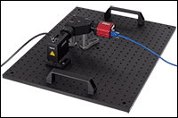
A full Raman spectroscopy system configured for solid samples. Included in the setup are a RSB1(/M) Base Unit, a RSBR1(/M) Reflective Front End (with the stray light cover), and an armored FC/PC fiber patch cable for a 785 nm source.
Click to Enlarge
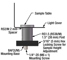
Sample Stage Components
- Designed for Use with Powder and Solid Samples
- FC/PC Fiber-Coupled Input for the Excitation Laser
- Height-Adjustable Sample Stage for Samples of Varying Thickness
- Includes Polystyrene Chip Calibration Sample
The RSBR1(/M) Reflective Front End is designed to optimize collection of Raman-scattered light from powder and solid samples. Back-scattered Raman signal collected from the sample is carried through an optical baffle assembly, eliminating unwanted stray light, and then focused onto the coded input aperture of the base unit. Intended for use with a fiber-coupled excitation source, this front end features a FC/PC connector and is designed for excitation between 680 nm and 785 nm. For 785 nm excitation, we recommend using the Thorlabs FPV785M VHG-Stabilized 785 nm Laser.
The main body of this front end, which includes a 680 nm to 785 nm bandpass filter, an 810 nm longpass filter, and collection mirror, connects to the input aperture of the base unit through the SM1 (Ø1.035"-40) internal threads on the optical baffle assembly. An SM1 locking ring is also included on the optical baffle, which has a factory-set position for ideal performance of the front end.
*Note that RSBR1(/M) front ends built in 2021 or 2022 accommodate excitation only at 785 nm. Please contact Tech Support for assistance if you plan on adapting older front ends for use with excitation between 680 nm and 785 nm.
Each reflective front end includes the main body and attached optical baffle, a height-adjustable sample table, and a stray light cover. Also shipped with each unit is a polystyrene chip calibration sample (Item # RPC); replacement calibration samples can be purchased below. For the full list of components shipped with this front end, please see the Shipping List tab.
Sample Stage
For best performance, the sample needs to be in the focal plane of the collection mirror, which is 2.08" (52.6 mm) or 54.1 mm (2.14") above the breadboard for imperial and metric front ends, respectively. The included sample stage should be positioned directly underneath the window of the RSBR1 front end and can be mounted to the breadboard using the counterbored slots in the included BA2F(/M) mounting base; secure the mounting screws with a 0.2" (5 mm) hex key.
The sample stage is shipped with the sample table at a height optimized for the included polystyrene calibration chip, however, this height is adjustable to accommodate samples of varying thicknesses. For thinner samples, the sample table can be raised by 7.0 mm (0.27") using the locking screw in the BA2F(/M) base and a 3/16" (5 mm) hex key. Thicker samples can be evaluated by removing the 2 mm spacer and 1.5" (38 mm) mounting post and replacing them with a shorter post.
Stray Light Cover
To prevent unwanted light from entering or exiting the setup, a 2-piece stray light cover is included with the reflective front end. The U-shaped bottom piece sits underneath the sample stage and fits around the lens tube of the optical baffle assembly, while the top cover fits around the main body of the front end and sample area. The top cover is secured into place using three 8-32 (M4) cap screws.
This light cover does not fulfill laser safety standards and can only be installed when the laser input port points upward. Also note that when paired with a user-supplied laser source, the front end provides a free space beam. Necessary laser safety protocols will depend on the user's chosen laser source and the lab environment. Please consult your laser safety officer to ensure the safety of the setup before powering on any laser source.
Note: Do not mix imperial base units with metric front ends or vice versa. The input aperture height between imperial and metric units differs slightly due to the mounting posts.

Must be Purchased Separately
- RSB1(/M) Base Unit
- Source with Excitation between 680 nm and 785 nm (See Laser Requirement Tab)
- PC for Running Thorlabs OSA Raman Software
For the full list of components shipped with this front end, please see the Shipping List tab.
Click to Enlarge
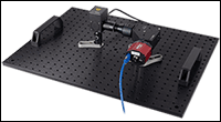
A full Raman spectroscopy system configured for liquid samples. Included in the setup are a RSB1(/M) base unit, a RSBC1(/M) cuvette front end, and an armored FC/PC fiber for a 785 nm source.
- Designed for Use with Transparent, Semi-Transparent, and Translucent Materials
- Adaptable for Polarization-Dependent Raman Measurements
- FC/PC Fiber-Coupled Input for the Excitation Laser
- Ideal for 3500 µL Fluorescence Cuvettes with a 10 mm Transmitted Path Length
- Polystyrene Block Calibration Sample
The RSBC1(/M) Cuvette Front End is designed for Raman spectroscopy measurements on transparent, semi-transparent, and translucent materials. Raman-scattered light is collected in a 90-degree geometry with respect to the excitation source, reducing the amount of stray laser light and Rayleigh-scattered photons that enter the spectrograph. Intended for use with a fiber-coupled excitation source, this front end features a FC/PC-terminated collimator and is designed for excitation between 680 nm and 785 nm. For 785 nm excitation, we recommend using the Thorlabs FPV785M VHG-Stabilized 785 nm Laser.
Each cuvette front end includes the main body, an optical baffle, a 3500 µL synthetic quartz glass cuvette, and a light-tight cover. Also shipped with each unit is a polystyrene block calibration sample (Item # RPB); replacement calibration samples can be purchased below. For the full list of components shipped with this front end, please see the Shipping List tab.
*Note that RSBC1(/M) front ends built in 2021 or 2022 accommodate excitation only at 785 nm. Please contact Tech Support for assistance if you plan on adapting older front ends for use with excitation between 680 nm and 785 nm.
The main body of this front end, which includes a CVH100 Cuvette Holder, collimator, a 680 nm to 785 nm bandpass filter, and an 810 nm longpass filter, connects to the input aperture of the base unit through SM1 (Ø1.035"-40) internal threads on the optical baffle assembly. The distance between the base unit and front end is critical for performance of the Raman kit, and an SM1 locking ring and SM1M05 connecting ring are included on the baffle to achieve the necessary 1.5 mm (0.59") separation. A detailed procedure for setting up this front end can be found in the user manual.
When paired with a user-supplied laser source, the front end collimates the laser for excitation. Necessary laser safety protocols will depend on the user's chosen laser source and the lab environment. Please consult your laser safety officer to ensure the safety of the setup before powering on any laser source.
| Compatible Cuvette Specifications | |
|---|---|
| Light Path | 10 mm |
| Dimensions | 12.5 mm x 12.5 mm x 45 mm (0.49" x 0.49" x 1.77") |
| Optical Axis Height Above Base |
8.5 mm |
| Polished Sides | 4 |
Cuvette
This front end ships with a 3500 µL synthetic quartz glass cuvette, which has four polished sides, a standard 12.5 mm square outside dimension, and a 10 mm transmitted path length through the sample. The cuvette is engraved with a letter "Q" that designates its synthetic quartz glass construction and the optical window axis, and it comes with a PTFE cap to prevent contamination by dust or other particles. Additional Synthetic Quartz Glass Cuvettes are also available for purchase separately, including the CV10Q35F Macro Fluorescence Cuvette which matches the specifications shown in the table to the left.
The cuvette included with this kit should not be used with benzene, toluene, aqua regia, ethanol, corrosive solutions, or other similar substances, as they may degrade the bonds between the pieces and cause the cuvette to leak.
Polarization-Dependent Measurements
Controlling the polarization of the excitation and Raman-scattered light can provide information about the symmetry of bond vibrations within a molecule. In the as-delivered configuration (90° geometry), this front end only detects s-polarized light unless the excitation light is completely unpolarized. The signal strength of peaks resulting from symmetrical vibrational modes can be optimized by rotating the collimator, which can be done by loosening the 1/16" (1.5 mm) nylon-tipped screws that secure the collimator in the fiber input. It is also possible to include polarization optics into the excitation and collection paths; please see the Polarization tab for more information.
Please note that it is possible to remove the collimator from the front end, which presents the risk of radiation exposure when connected to a user-supplied laser. Necessary laser safety protocols will depend on the user's chosen laser source and the lab environment. Please consult your laser safety officer to ensure the safety of the setup before powering on any laser source.
Note: Do not mix imperial base units with metric front ends or vice versa. The input aperture height between imperial and metric units differs slightly due to the mounting posts.

Click to Enlarge
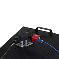
The RSBF2 Fiber Input Adapter couples Raman-scattered light collected with an RP35 Fiber Probe (sold below) from user-designed setups into the RSB1(/M) base units.
Click to Enlarge
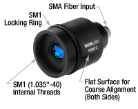
RSBF2 Adapter Housing Features
- Couples Fiber-Coupled Raman-Scattered Light to the RSB1(/M) Base Unit
- Optimized for Raman Signal in the 650 nm to 1050 nm Wavelength Range
- SMA Fiber Input Port
The RSBF2 Fiber Input Adapter allows fiber-coupled Raman signal from a user-designed setup to be coupled into the RSB1(/M) base unit. Designed around a fly’s eye homogenizer, this adapter transforms the output of an optical fiber, which is a round beam with a Gaussian profile, into a rectangular homogenized spot that matches the shape and evenly illuminates the base unit’s coded input aperture. This adapter is designed for Raman-scattered light from 650 nm to 1050 nm and is compatible with both the imperial and metric base units.
The main body of this adapter includes three main components: an F240SMA-850 fixed-focus fiber collimator with an SMA905 connector, a fly’s eye homogenizer for generating a square beam, and a lens for focusing the square beam onto the input aperture of the base unit. The housing connects to the input aperture of the base unit through SM1 (Ø1.035"-40) internal threads. For best performance, we recommend using SMA-terminated multimode fiber patch cables with a core diameter <2.5 mm and NA <0.5, such as our RP35 Raman Fiber Probe (sold below) or our M29L01 and M93L01 patch cables.
A flat edge on the housing and a locking ring can be used for coarse and fine alignment of the square beam to the input aperture, respectively (see image above). When coarsely aligned, the flat edge of the adapter housing is parallel to the base unit. The locking ring can then be loosened, which allows the adapter body to rotate for fine adjustments to the alignment. We recommend using a M850F3 850 nm Fiber-Coupled LED and our ThorCam™ software to image the resulting rectangular beam shape and confirm good alignment of the system; see the manual for a detailed alignment procedure and the Software tab for more information on ThorCam.


Click to Enlarge
A schematic of the RP35 Fiber Probe fiber configuration. The light source and spectrometer legs are also referred to as excitation and emission legs, respectively.
- Fiber Y-Cable Ideal for use wih Solid, Liquid, and Powder Samples
- Designed for a 680 - 785 nm Excitation and an 815 - 950 nm Detection Wavelength Range
- SMA Output Connector for Compatibility with the RSBF2 Fiber Input Adapter
The RP35 Reflection Probe for Raman Spectroscopy is a fiber optic Y-cable optimized for measuring 815 - 950 nm Raman-scattered light from solid, liquid, and powder samples. Made from Ø200 µm Low-OH silica fibers, this fiber probe has a leg to carry light from a light source to a sample and a leg to carry light scattered by the sample to a spectrometer. Please note that this fiber probe is designed to be used with the RSBF2 Fiber Adapter (sold above); the adapter transforms the round output profile from the probe into a rectangular spot that matches the shape of coded aperture used in the RSB1(/M) Base Unit.
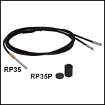
The light source leg of this probe features a single fiber and includes an FC/PC connector, which provides compatibility with fiber-coupled laser sources such as our FPV785M VHG-Stabilized 785 nm Laser. For use with a variety of sample types, the sample leg has a Ø1/4" probe consisting of a single fiber, which delivers excitation light to the sample, that is surrounded by 67 low-OH silica fibers (maximum of two broken) for collecting Raman signal from the sample. To widen the field of view, a fused silica ball lens with a 1.0 mm working distance is located at the tip of the probe. A short pass and long pass filter are included in the sample end probe, which block Raman-scattered light (generated within the fibers) from reaching the sample during excitation and prevent the collection of any scattered laser light, respectively. The spectrometer leg includes an SMA905 connector for compatibility with the RSBF2 Fiber Input Adapter and has a round bundle of 67 fibers that provides a Ø2.1 mm illumination area. See the image to the right for detailed views of the probe leg end-faces.
Please note that we recommend having the probe in contact with the sample or keeping the distance between the tip and sample <0.5 mm. For mounting the probe in custom setups, the RPS Fiber Optic Probe Stand can be used to firmly secure the probe at adjustable heights above a sample.
Three plastic caps are included with this probe to protect the fiber tip and fiber connectors. Also available is the optional SM05-threaded RP35P Protective Cap which contains a polystyrene sample (Rexolite®* 1422) for calibrating the spectrometer and confirming the wavelength of the excitation laser.
Laser Warning
The RP35 Fiber Probe for Raman Spectroscopy has the same laser class as the excitation laser used, so it is assumed to be a class 4 laser device in most cases. For further laser safety information, please refer to the user manual, which can be found by clicking on the red Docs icon (![]() ) next to the Item #.
) next to the Item #.
*Rexolite® is a registered trademark of C-Lec Plastics Inc.
| Item #a | Hydroxyl Content |
Wavelength Range | Fiber Type | Light Source Legb | Sample Leg | Spectrometer Legc | Fiber Core Diameter | NA | Minimum Bend Radius |
|---|---|---|---|---|---|---|---|---|---|
| RP35 | Low-OH | 680 - 785 nm (Excitation) 815 - 950 nm (Collection)d |
Fused Silica | FC/PC Narrow Key Connector, Single Fiber |
Ø1/4" Probee |
SMA Connector, 67-Fiber Round Bundle |
200 ± 4 µm | 0.22 ± 0.02 | 80 mm |

Click to Enlarge
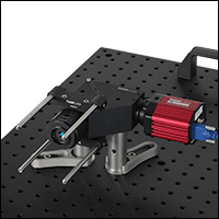
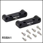
The RSBA1 Adapter provides compatibility with 60 mm cage system components.
- Provides Compatibility with Thorlabs' 60 mm Cage System Components
- Ideal for Attaching Custom-Built Front Ends to the RSB1(/M) Base Unit
- Each Adapter Includes:
- Two Brackets
- Four 8-32 Cap Screws (9/64" Hex)
- Four M4 Cap Screws (3 mm Hex)
The RSBA1 60 mm Cage System Adapter provides an option for applications that require a custom-built front end for the base unit input aperture. Each adapter consists of two brackets that can be attached to an RSB1(/M) base unit using the included cap screws. Two dowel pins on each bracket fit into holes on the base unit to aid with accurate alignment. Each bracket also features two through holes for our Ø6 mm cage system construction rods; these are spaced apart by 60 mm for compatibility with our 60 mm cage system components. The cage rods can be locked into place using a 5/64" (2 mm) hex key.

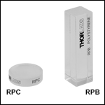
- RPC: Replacement Polystyrene Chip for the RSBR1(/M) Reflective Front End
- RPB: Replacement Polystyrene Block for the RSBC1(/M) Cuvette Front End
The RPC polystyrene chip and RSB polystyrene block are replacement calibration samples for use with the Raman spectroscopy kit front ends.

| Key Specifications | |
|---|---|
| SERS Active Material | Gold |
| Active Area |
4.3 mm x 4.3 mm |
| Laser Wavelength (Recommended) | 785 nm |
| Minimum Power Densitya (Recommended) |
0.1 W/cm2 |
| Lifetimeb (Recommended) | <6 Months |
| Dimensions (L x W x H) | 4.5 mm x 4.5 mm x 0.5 mm (0.18" x 0.18" x 0.02") |
- Gold-Coated Textured Fused Quartz Substrate
- Dimensions: 4.5 mm x 4.5 mm x 0.5 mm
- Active Area: 4.3 mm x 4.3 mm
- Optimized for 785 nm Laser Excitation
- Compatible with Thorlabs' Modular Raman Spectroscopy Kit and Portable Raman Spectrometer
- Available in 5 Pack
The RCT4M Surface-Enhanced Raman Spectroscopy Substrate is designed for use in SERS to enhance Raman scattered signal by orders of magnitude. The gold-coated textured quartz substrate enables sensitivity down to parts per billion,* allowing for the measurement of low sample concentrations with significantly lower laser powers in comparison to standard Raman spectroscopy.
The SERS substrate is optimized for 785 nm laser excitation and is compatible with Thorlabs' Modular Raman Spectroscopy Kit. To avoid damaging the plasmonic nanostructures and contaminating instrumentation, ensure the active surface of the substrate does not make direct contact with the mounting surface. Please note that the SERS substrate is single-use and washing is not recommended.
The SERS substrate is packaged inside a vacuum sealed bag. For best performance, it is recommended to use the substrate within 6 months when outside of vacuum. For more information, please see our full presentation on Surface-Enhanced Raman Spectroscopy Substrate.
*Sensitivity is dependent on the experimental conditions, including the analyte, incident laser power, and integration time.
 Products Home



 Zoom
Zoom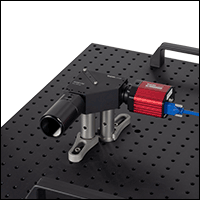

 Modular Raman Spectroscopy Kit